Focus on your quality of life
Being pain-free and mobile is the basis for quality of life. Dr. Olischar offers a wide range of non-surgical therapies for this purpose. They are just as much part of his core competencies as endoprosthetics and arthroscopy for surgical procedures. The use of modern C-arm-technology for targeted pain therapies rounds out the special offering for state-of-the-art, efficient treatments. Physiosteps²’s first-class physiotherapy offers additional options for achieving optimal results.
Dictionary
The lexicon describes the therapies and their areas of application as briefly and clearly as possible.
AC joint resection - coplaning
AC joint resection – Coplaning:
Used totreat acromioclavicular joint osteoarthritis. Through arthroscopic surgery, the outer end of the clavicle is removed with a small burr along with the inflamed bursa below the acromion. Thus, more space is created in the subacromial space for smooth gliding of the tendons.
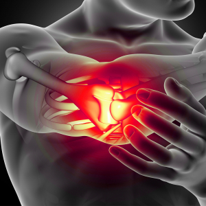
3D render of a male figure holding elbow in pain with close up of elbow bone
ACP Treatment (Autologous Conditioned Plasma)
ACP Treatment (Autologous Conditioned Plasma):
During healing, complex and precisely regulated natural processes take place in the body. Special proteins, the so-called growth factors, which originate from small blood components, the thrombocytes – blood platelets, are involved in this. These are activated in the body at the site of the injury and release growth factors there, which in turn promote the healing process. The procedure is as follows:
- blood sampling from the arm vein
- separation procedure to obtain the body’s own active substances (proteins/growth factors) in concentrated form.
- injection of the body’s own active substances into the affected region – site of injury
ACP Treatment is based on this principle. ACP Treatment is based on this principle. Here, the body’s own self-healing powers are used by obtaining high concentrations of the special proteins/growth factors and then injecting them. Depending on the type of injury, the form and frequency of administration of these proteins can vary.
An individual treatment plan is designed with your doctor, e.g. several injections at weekly intervals.
Arthrodesis joint fusion
Arthrodesis joint fusion:
Arthrodesis is a surgical fusion of a joint. The ability to move in the joint is thereby completely suppressed, resulting in freedom from pain. If only temporary fixation is applied across a joint, e.g. with a K-wire, a temporary arthrodesis is present.
In principle, surgical joint fusion is possible on all joints. Hip and knee arthrodeses have become rare since the introduction of arthroplasty. Relatively often, the procedure is still performed on the shoulder, wrist and finger joints, ankle joints and ankles. Arthrodesis of the first tarsometatarsal joint (lapidus arthrodesis) is a common procedure to correct severe unstable hallux valgus, and metatarsophalangeal joint arthrodesis is often performed for hallux rigidus.
Arthroscopy
Arthroscopy:
With the help of arthroscopy or joint endoscopy, larger joints can be examined and damage to the joint contours can be treated. For this purpose, a so-called arthroscope is inserted through a small incision in the skin. This is a rod-shaped video camera. For a better assessment of the joint structures, a light source and a rinsing and suction device are also attached. Furthermore, special instruments can be used arthroscopically so that damage and injuries can be treated immediately after their diagnosis. Arthrosis:
Arthrosis is the typical sign of wear and tear on the joints and a natural consequence of our ageing process, but arthrosis can also occur in younger people (due to accidents, sports, or congenital conditions).
Osteoarthritis is a wear and tear of the articular cartilage. The destruction of the joint ultimately leads to a painful restriction of movement and painful bone overlay. Treatment depends on the stage of the arthrosis.
The first signs / symptoms of arthrosis are:
- Starting pain and joint stiffness
- Restricted mobility
- Rest and night pain
- Inflammation and swelling
Arthrosis is divided into stagesfromI to IV :
Stage I: Symptomless Stage II – III: Increase in pain Stage IV: Complete wear and tear of the joint, restriction of movement
Arthroscopy of the ankle joint
Arthroscopy of the ankle joint:
Arthroscopy, or reflection, is a joint-preserving procedure on the ankle joint. The surgical procedure is performed using a minimally invasive technique, which significantly reduces healing times, scarring and complications compared to open surgery. Minimal skin incisions means less tissue trauma for the patient and therefore less pain.
The following therapies can be performed during ankle arthroscopy:
- Treatment of cartilage damage in the ankle joint
- Removal of free joint bodies in the ankle joint
- Treatment of cartilage damage
- Diseases of the synovial membrane (synovitis) with entrapments
- Treatment of impingement in the ankle joint
- Treatment of instabilities of the ankle joint (lateral ligaments, syndesmosis)
- Osteoarthritis and its sequelae of the ankle joint
Arthroscopy of the hip joint
Arthroscopy of the hip joint:
Hip arthroscopy or endoscopy of the hip joint is a procedure in which the joint space between the femoral head and the acetabulum as well as the rest of the joint interior can be examined. The patient receives general anesthesia for the surgical procedure
The following therapies can be performed as part of hip arthroscopy:
- Removal of free joint bodies from the hip joint
- Treatment of cartilage damage in the hip joint
- Treatment of diseases of the synovial membrane of the hip joint
- Treatment of shape disorders of the hip joint (CAM and pincer impingement of the hip)
- Therapies for damage to the joint lip: injuries to the labrum
Arthroscopy of the shoulder joint
Arthroscopy of the shoulder joint:
Arthroscopy of the shoulder joint is a minimally invasive surgical procedure to better evaluate the cause of pain and simultaneously treat it. The keyhole technique causes minor muscle damage, which in turn leads to a reduction in shoulder pain, resulting in better mobility.
- Subacromial decompression:
Subacromial decompression (S.A.D.) is a surgical procedure used to treat impingement syndrome. In this case, there is an obstruction of the supraspinatus tendon as it slides under the acromion. Inflammatory processes, bone pull-outs at the acromioclavicular joint, a pronounced AC joint arthrosis or calcified shoulder,can be responsible for this. The inflamed bursa below the acromion is removed. If a bone spur is present on the underside of the acromion, it is removed with a bur to create a flat surface. The space between the clavicle and the acromion is widened so that the tendons that slide underneath can work smoothly. - AC joint resection (Coplaning):
Used for the treatment of acromioclavicular joint osteoarthritis. Using arthroscopic surgery, the outer end of the clavicle is removed with a small burr along with the inflamed bursa below the acromion. Thus, more space is created in the subacromial space for smooth gliding of the tendons. - Suture of the rotator cuff:
Basically, tendons connect the associated muscle to a specific bone, providing specific movement. The rotator cuff is 4 interrelated muscles that are responsible for inward and outward rotation of the upper arm, stabilizing the arm to the torso, and splaying and overhead lifting of the arm. Increasing wear as a result of high mechanical stress or accidents can lead to a tear in one or more tendons of the rotator cuff. RM suture is a suture of one or more torn or injured tendons with the goal of regaining full strength and mobility and eliminating pain. - Tenotomy u./u. Tenodesis of the long biceps tendon:
The long biceps tendon is a problem child of the shoulder joint. Both in the area of its attachment to the supraglenoid tuberosity (SLAP lesion) and in its intra-articular course up to its entry into the sulcus (Pulley lesion), there is structural damage that can lead to considerable pain. This results in functional and movement restrictions. In the rarest cases, anatomical reconstruction is useful in these pathologies. Usually, the question of extraanatomical fixation (tenodesis) or simple detachment (tenotomy) of the long biceps tendon arises. - Removal of calcium deposits:
Calcium shoulder is the term used to describe tendon calcification (tendinosis calcaria) in the shoulder joint. It occurs mainly between the ages of 30 and 50. The courses are very different. In most cases, pain therapy and physiotherapy alleviate the symptoms; in rare cases, surgery is necessary. This is done arthroscopically, the so-called keyhole method. The camera and instruments are inserted into the subacromial space through small skin incisions and the calcium deposit is visited. Subsequently, the calcium focus is peeled out of the tendon. If larger defects occur in the tendon, these are treated with sutures.
Bandaging– orthopedic technology
Bandagisterei – Orthopedic technology:
The Bandagist (orthopaedic technician) manufactures custom-made orthopedic/technical aids for patients according to a doctor’s prescription. These are used for the conservative treatment of orthopedic problems and damage to the postural and musculoskeletal system, joint misalignments and instabilities. This includes prostheses and orthoses.
In addition, orthopedic technology mechanics manufacture special bandages and corsets, but also rehabilitation technology products such as seat shells or individual wheelchairs. Depending on the aid, fittings are already carried out during the manufacturing process in order to check the position of the aid and correct it if necessary. They advise their customers on the choice of the right aid, carry out anamnesis, measure affected body parts, create construction drawings and models.
In addition, they repair, maintain and adjust the tools and instruct them in their operation and handling. The orthopedic technology trade belongs to the health professions and to the group of health trades.
Bursitis
Bursitis:
When a bursa becomes inflamed, more fluid than normal accumulates inside it. Experts call this an effusion. This causes a swelling that can be felt and easily seen from the outside, especially if the inflamed bursa is located close under the skin.
Bursitis usually develops after excessive physical exertion. Characteristic is sudden onset of severe pain, usually first appearing at night after the overload. A bacterial infection can also lead to the clinical picture. Wear and tear and chronic overuse also sometimes cause bursitis.
Treatment (conservative): Immobilization of the affected joint, analgesics, sometimes cortisone, shock wave therapy, puncture to aspirate excess fluid, physical therapy, antibiotic administration if necessary.
Bacterial or chronic bronchitis often requires surgical removal.
C-Arm technology
C-arm:
The C-arm is a medical device for imaging. Characteristic is the C-shaped design, with which the patient can be examined from almost any angle. The C-arm solved the problem of immobility, reduces strain as well as waiting time, and offers the option to move the device to the patient as needed.
The C-arm is particularly suitable for intraoperative imaging. Here, every step of the operation can be monitored in real time, so that a revision can be made if necessary and a possible follow-up operation can be saved, treatment results improved and recovery accelerated.
Imaging and CT-targeted infiltration procedures have become indispensable in modern pain therapy, especially in the spine.
Thanks to imaging, highly selective anesthesia is used to place the active ingredients at the site of the pain and thus eliminate the cause of the pain. Therapeutically, these infiltration methods can also be used to excellent effect and in some cases can be complemented by more advanced interventional procedures such as denervation.
Calcareous deposit - Tendinitis calcarea
Calcareous tendinitis – Tendinitis calcarea:
Calcareous shoulder is the term used to describe tendon calcification (tendinosis calcaria) in the shoulder joint. It occurs mainly between the ages of 30 and 50. The courses are very different. In most cases, pain therapy and physiotherapy alleviate the symptoms; in rare cases, surgery is necessary. Basically, tendons connect the associated muscle to a specific bone and thus provide a specific movement. The calcified shoulder mainly affects the tendons of the rotator cuff:
- Supraspinatus tendon: Most commonly affected, allows the arm to splay out
- Infraspinatus tendon: Frequently affected, is responsible for external rotation of the arm
- Subscapularis tendon: Less frequently affected, serves for internal rotation of the arm
Both conservative procedures (shock wave, needling, antiphlogistic therapy, physiotherapy) and surgical procedures (arthroscopic calcific foci removal) are used in the treatment of calcific shoulder.
Cam and Pincer - Impingement
CAM and PINCER Impingement – Femoro-Acetabular Impingement (F.A.I.):
Impingement syndrome of the hip refers to a mechanical tightness between the head of the femur and the acetabular roof. Bony changes of the femoral neck or the acetabular roof cause painful banging of both joint partners. Mostly young, athletically active people are affected by impingement syndrome of the hip. The pain manifests as motion-dependent groin pain, pain when standing up after prolonged sitting, resulting in limited mobility in the hip joint.
With timely therapy, it may be possible to prevent more severe joint damage (arthroscopic surgery); without therapy, the cartilage or joint lip will continue to be damaged, which can lead to hip joint arthrosis and, in the worst case, artificial hip replacement.
Carpal-tunnel-syndrome
Carpal tunnel syndrome:
Carpal tunnel syndrome is a constriction of the median nerve in the wrist tunnel (carpal tunnel). This channel for nerves and tendons is formed in a U-shape by the carpal bones and covered by a band of connective tissue (retinaculum flexorum). This creates a bottleneck in the tendon compartment of the wrist. This pinches the median arm nerve, which supplies sensory and motor functions to various parts of the hand. Typical symptoms include the hand falling asleep at night, feelings of numbness and insensitivity, pain, and later even paralysis and functional disorders. Treatment is primarily aimed at immobilizing the wrist at night using an orthosis, cortisone treatment and, if necessary, surgical widening of the carpal tunnel.
Cartilage: Osteochondral injuries
Osteochondral Injuries:
An osteochondral lesion is a combined damage to the cartilaginous sliding layer as well as the underlying bony bedrock. The discomfort is usually felt in the depth of the joint during weight bearing. The patient suffers from severe pain in the affected joint and limited mobility. These injuries may be traumatic or caused by osteochondrosis.
Osteochondrosis is a wear-related (degenerative) disease of cartilage and bone in the joints. This means that both cartilage and bone are involved in the disease process.
Osteochondrosis dissecans represents a special form. This results in the death of small areas of bone below the articular cartilage in the joint. This disease usually occurs in children and adolescents aged 5-20 years. Early recognition and treatment of osteochondrosis dissecans is important. If the disease progresses untreated, the joint cartilage also suffers damage. The result can be premature joint wear (arthrosis) with chronic complaints and functional disorders of the joint.
Arthroscopic surgery (using the keyhole technique) can remove the damaged portions of cartilage and bone and stimulate the formation of regenerative tissue. Other surgical procedures such as mosaicplasty, microfracturing or even cartilage transplantation are used here.
Cartilage formation / Cartilage damage
Cartilage development – hyaluronic acid therapy:
Artificial synovial fluid – Our joints produce the synovial fluid (joint fluid) for smooth movement.
- Lubricant: friction is reduced
- Shock absorber: it buffers shocks, (e.g. jumps)
- Placeholder: it keeps the joint surfaces at a distance (sliding, riding up)
- Filter function: it filters out harmful substances and supplies the cartilage with nutrients
Claw toe
Claw toe – claw toe – hammer toe:
These are malpositions of the toes with a flexion in the middle joint and an alternating position in the base and end joint. The pressure of the bone under the skin leads to the formation of painful pressure points, so-called calluses, and to bursitis over the affected joints. Hammer toe is usually accompanied by a deformity of the ball of the foot.
The surgical treatment of hammer toe involves removing the head of the proximal phalanx. This allows the misposition to be corrected. To maintain the result of the correction, it is stabilized with a wire (temporary joint stiffening), which is usually removed again painlessly after 4-6 weeks.
Coplaning - AC joint resection
Coplaning – AC Joint Resection:
Used for the treatment of acromioclavicular joint osteoarthritis. Through arthroscopic surgery, the outer end of the clavicle is removed with a small burr along with the inflamed bursa below the acromion. Thus, more space is created in the subacromial space for smooth gliding of the tendons.
Coxitis fugax
Coxitis fugax– hip flare:
coxitis fugax is a non-infectious inflammation of the joint with spontaneous healing. It occurs more frequently between the ages of 4 and 10. Often, coxitis fugax is preceded 1-3 weeks earlier by a (viral) infection of the respiratory tract or gastrointestinal tract. Patients express sudden pain that often radiates to the knee. Affected children limp to protect the affected leg. The general condition of affected children is usually good and stable. Examination of the hip reveals a painful restriction of motion, particularly emphasized during internal rotation.
Therapy included rest for 2-4 days and possible administration of anti-inflammatories/NSAIDs (e.g., ibuprofen). Ultrasound-targeted follow-up and control of laboratory parameters (inflammatory values) are important. If the effusion is pronounced, the joint can be punctured for diagnostic and therapeutic purposes.
Cruciate ligaments
Cruciate ligaments:
Definition: The cruciate ligaments form the central passive guiding elements of the knee joint. Due to their position relative to each other and their type of fixation on the femur and tibia, they form a locking chain and hold/stabilize the femoral head during flexion in a roll-slip mechanism roll-slip mechanismthat allows a large condyle to move physiologically on a much smaller socket. Function: The cruciate ligaments together with the collateral ligaments hold the knee joint together. They limit the extension of the tibia, guide the joint during movement and give it the necessary stability. If the anterior cruciate ligament is damaged, this is severely disturbed and leads to cartilage and meniscus damage.
Degenerative joint wear
Degenerative joint wear:
Signs of wear or abrasion cause pain-related functional limitations. Osteoarthritis is a progressive breakdown of the shock-absorbing cartilage layer between two joint surfaces. Osteoarthritis patients suffer from severe joint pain and increasingly limited mobility. In the advanced stage, stiffening of the joint is imminent. With appropriate treatment as early as possible, arthritic symptoms can be alleviated or even eliminated.
Disc prolapse
Herniated disc:
A herniated disc (disc herniation, disc prolapse) most commonly occurs in people between the ages of 30 and 50. This causes severe back pain, numbness and even paralysis.
Here, the soft nucleus (nucleus pulposus) emerges from the disc located between 2 adjacent vertebrae. It is usually located inside a solid fibrous (annulus fibrosus) that is damaged or unstable in a herniated disc. This can cause the nucleus to bulge out of the disc or even pass through the annulus. In pronounced cases, parts of the gelatinous nucleus can slip into the spinal canal, a so-called sequestered disc herniation. If the nucleus protrudes from the intervertebral disc, this often causes the nerves emerging between the vertebrae (spinal nerves) or the spinal cord lying in the spinal canal to be compressed. Pain and dysfunction of the spinal cord occur.
The herniated disc (disc prolapse) must be distinguished from the bulging disc (disc protrusion). Here, the inner disc tissue shifts outward without rupturing the fibrous ring of the disc. Nevertheless, complaints such as pain and sensory disturbances may occur. A well-known example is lumbago, an acute, sharp pain in the area of the lumbar vertebrae.
Dupuytren’s disease
Dupuytren’s disease:
Dupuytren’s disease is a benign disease of the connective tissue of the palm. In this case, a benign tumor forms, which over time impedes the extension of the affected fingers. Characteristic of the disease is the appearance of nodules, connective tissue hardening (hard swellings) and strands on the inner surface of the hand. The growing strands can eventually lead to an inability to extend the finger (e.g., retractions with stiffening, extension deficit, flexion contracture). The base and middle joints of the fingers are particularly affected.
If one or more fingers are curved and cause a troublesome disability, appropriate surgery can usually restore finger extension. Conservative therapy by means of irradiation or shock wave is suitable for this purpose. In the absence of success, it is necessary to think about surgical removal and solution of the strands. In case of a surgical procedure, it is important to point out the high recurrence rate of the nodes.
Men are affected 5 times more often than women.
Endoprosthetics - hip, knee
Endoprosthetics – hip, knee
In orthopedic surgery, endoprosthetics refers to the field of “artificial joint replacement”.
Our musculoskeletal system consists of a complex assembly of different structures that enable us to move purposefully.
Osteoarthritis is a wear and tear of the articular cartilage. The destruction of the joint in leads to a painful restriction of movement and a painful build-up of bone (exostoses, osteophytes). Treatment depends on the stage of the arthrosis. If the arthrosis is far advanced, joint preservation is not possible. In this case, depending on the localization and severity of the joint degeneration, the only option is an osteotomy or an artificial joint replacement.
Epicondylitis: tennis elbow and golfer’s elbow
Epicondylitis (tennis elbow and golfer’s elbow):
Epicondylitis describes a painful irritation of the tendon origin on the inside, or outside, of the elbow. The muscles of the forearm above the epicondyle (bony prominence) on the humerus are affected. The disease associated with the group of enthesiopathies.
Epicondylitis radialis humeri-Tennisellenbogen: at the outer epicondyle of the humerus – extensor of the wrists and fingers
Epicondylitis ulnaris humeri-buyer elbow:at the inner epicondyle of the humerus-bend of the wrist and fingers
Femoral head necrosis
Femoral head necrosis:
in femoral head necrosis, part of the bony head of the femur dies. The disease belongs to the aseptic bone necrosis. The cause is reduced blood flow leading to necrosis. A special case of femoral head necrosis is Perthes disease in childhood. In adults, there is often an association in diabetes mellitus, alcoholism, and after high-dose cortisone treatment and prolonged treatment with anticoagulants.
Frozen Shoulder
Frozen Shoulder – Adhesive Capsulitis:
Fibrosis shoulder refers to a disease of the capsule of the shoulder joint. Initially, there is severe shoulder pain, which gradually subsides, while at the same time the shoulder becomes increasingly stiff. Here will be divided into 3 phases. In the first phase, severe shoulder pain occurs sometimes at rest and at night. During the second phase, the shoulder joint stiffens more and more and in phase three, a reversal of the symptoms takes place, the mobility of the shoulder increases again. Sometimes protracted courses over several years are predicted, sometimes without complete healing and long-term restriction of movement.
The therapy provides for ice or heat treatments, physiotherapeutic exercise treatments and/or exercise baths, antiphlogistic therapy with anti-inflammatory drugs, sometimes in combination with cortisone administration.
Gout / gouty thophus - arthritis urica
Gout / Goutthophus – Arthritis urica:
this is an inflammatory nodule formation whereby it comes to a tissue deposition of uric acid in the subcutis and other connective tissue structures.
- Conservative: The preferred treatment for gout nodules today is medication to lower uric acid levels. If the uric acid level is below 6.0 mg/dl, the nodules break down again on their own. The lower the uric acid level, the faster the gout nodule shrinkage. Therefore, a target value of less than 5.0 mg/dl uric acid p is aimed for in gout nodes.
- Operative: In case of an imminent tendon rupture, an operative procedure is recommended. Other indications for surgical intervention are constriction syndromes such as carpal tunnel syndrome or when necrosis is imminent.
Haglund’s exostosis
Plantar heel spur and Haglund’s exostosis:
Plantar Heel Spur:
A heel spur is a thorn-like ossification on the heel that is a few millimeters in size. Most often this results from irritation to the bone caused by traction or compression. This thorn-like bone block on the calcaneus is caused by small tendon injuries (microtrauma) that calcified. The reason for such injuries is usually misuse and overuse. With a heel spur, walking or standing is very painful in many cases.
Treatment: shoe inserts, cold therapy, physiotherapy, shock wave therapy, radiation, anti-inflammatory drugs, surgery.
The heel spur mainly occurs on the heel bone and can develop in the area of the Achilles tendon (dorsal – plantar fasciitis) or on the sole of the foot (plantar).Haglund’s exostosis is a special form.
Haglund’s exostosis:
Haglund’s exostosis describes an irritation of the Achilles tendon at the base of the calcaneus, which is usually combined with bursitis. In this case, there is a bony hump in the area where the Achilles tendon attaches, which causes the mechanical irritation and thus inflammation. Only adults are affected and the pain is located at the back of the heel and around the Achilles tendon insertion. Because the calcaneus forms a bone nose or protrusion at its posterior superior corner, Haglund’s exostosis is often called a posterior (dorsal) calcaneal spur.
If conservative therapy fails, surgical treatment is recommended.
Hallux rigidus
Hallux rigidus:
Hallux rigidus is a more advanced stage of arthrosis. The wear of the joint cartilage leads to a progressive destruction of the metatarsophalangeal joint with bony marginal attachments, which are responsible for the painful restriction of movement. Depending on the stage of the arthrosis, both conservative and surgical (joint-preserving or non-joint-preserving) therapy are available.
Hallux valgus
Hallux valgus:
The most common problems in the forefoot occur in the big toe. The bunion of the big toe or hallux valgus is defined as a deviation of the big toe at the base joint in the direction of the little toe. The incorrect loading of the foot leads to a splaying between the first 1st and 2nd metatarsal bones and thus to the emergence of the first metatarsal head, which forms the so-called bunion. A painful hallux valgus can usually only be treated surgically due to its malposition, considering the classification into mild, moderate, or severe form.
Hammer toe
Hammer toe – Claw toe – Claw toe:
These are malpositions of the toes with a flexion in the middle joint and an alternating position in the base and end joint. The pressure of the bone under the skin leads to the formation of painful pressure points, so-called calluses, and to bursitis over the affected joints. Hammer toe is usually accompanied by a deformity of the ball of the foot.
The surgical treatment of hammer toe involves removing the head of the proximal phalanx. This allows the misposition to be corrected. To maintain the result of the correction, it is stabilized with a wire (temporary joint stiffening), which is usually removed again painlessly after 4-6 weeks.
Heel spur - plantar or dorsal
Heel spur – plantar or dorsal:
A heel spur is a thorn-like ossification on the heel that is a few millimeters in size. Most often this results from irritation to the bone caused by traction or compression. This thorn-like bone block on the calcaneus is caused by small tendon injuries (microtrauma) that calcified. The reason for such injuries is usually misuse and overuse. With a heel spur, walking or standing is very painful in many cases.
Treatment: shoe inserts, cold therapy, physiotherapy, shock wave therapy, radiation, anti-inflammatory drugs, surgery.
The heel spur mainly occurs on the heel bone and can develop in the area of the Achilles tendon (dorsal – plantar fasciitis) or on the sole of the foot (plantar).Haglund’s exostosis is a special form.
Hip dysplasia
Hip dysplasia:
Hip dysplasia also called congenital hip dislocation, is a collective term for congenital or acquired deformities and disorders of ossification of the hip joint in the newborn. Hip dysplasia can occur alone or together with other congenital malformations. Solitary hip dysplasia is significantly more common and is found in significantly higher numbers in girls than in boys. Several factors are considered to favor or partially cause it: one factor is pelvic end presentation during pregnancy.
The symptoms of hip dysplasia are initially lateral imbalance of the gluteal folds and restricted movement of the affected hip during kicking. Without treatment, severe forms result in permanent damage to the hip joint with limping, gait disturbances and pain. The final state of severe forms is hip joint arthrosis. Mild forms do not exhibit pain. The presence of hip dysplasia is confirmed by using mainly sonography, in rarer cases X-ray and magnetic resonance or computed tomography. In most cases, treatment is carried out without surgery by using flexor-spreader orthoses (Tübinger splint, Pavlik bandage). Immobilization by application of a spreader cast (according to Ortolani) may also be necessary.
The need for surgical intervention is rare. The prognosis of hip dysplasia has improved considerably since the introduction of sonographic screening at neonatal examination.
Hyaluronic Acid Therapy
Hyaluronic acid therapy – cartilage regeneration:
Artificial joint lubricant – Our joints produce synovial fluid (= joint lubricant) for smooth running / movement.
- Lubricant: friction is reduced
- Shock absorber: it buffers shocks, (e.g. jumps)
- Placeholder: it keeps the joint surfaces at a distance (sliding, riding up)
- Filter function: it filters out harmful substances and supplies the cartilage with nutrients
Infiltrations - epidural sacral under C-Arm X-ray control
Infiltration – epidural, sacral under C-arm:
Epidural infiltration is the injection of drugs (local anesthetics, glucocorticoids) into the epidural space of the spinal cord, i.e. the space between the periosteum (bone skin) of the spinal canal and the hard meninges (dura mater).
Jumpers Knee
Jumper’s Knee (Patellar Pointe Syndrome):
Patellar Pointe Syndrome is a painful irritation condition at the lower pole of the kneecap. The disease is caused by an overload of the patellar tendon at the tendon insertion. It often affects athletes who perform jumping movements very often (e.g. volleyball, basketball, handball, long jump, high jump), hence the English name jumper’s knee.
With appropriate sports activity and load, pain occurs at the lower pole of the patella. Initially, these show up as start-up pain. As the inflammation in the tendon insertion area progresses, the pain also occurs in everyday life when the knee joint is extended against resistance (e.g. climbing stairs).
Loge de Guyon Syndrom
Loge de Guyon Syndrome:
Loge-du-Guyon syndrome is a nerve compression syndrome of the inferior segment of the ulnar nerve at the wrist. The Guyon’s loge is an anatomical constriction in the course of the ulnar nerve in the wrist region, which is bordered by two carpal bones as well as the retinaculum flexorum and musculus palmaris brevis. Due to the narrowness of the osteofibrous canal, mechanical overloading of the wrist (use of forearm crutches or cycling) may result in compression of the ulnar nerve with numbness. The paresthesias occur mainly in the area of the little finger.
The therapy depends on the cause of the complaints. Conservative therapy is to protect the nerve by immobilizing the wrist or avoiding overloading. A surgical procedure to relieve the nerve, especially in the case of mechanical constriction, is subsequently the treatment of choice.
Manual Therapy
Manual therapy:
Manual therapy is a treatment technique that addresses functional disorders of the musculoskeletal system (muscles and joints). It is applied in the spine and the joints of the extremities. Blocked or restricted joints often cause pain and tension in the muscles. These are mobilized with special hand movements and techniques, thereby increasing mobility, and reducing pain.
Mother-child examination: Hip ultrasound examination according to Graf in the 1st week of life
The importance of hip ultrasonography today lies not only in the early diagnosis of dislocation, but also, if possible, in prevention, i.e. to diagnose the joint at risk of dislocation on the right side and to prevent it from slipping into dislocation by means of sonography-guided therapy.
This examination detects congenital or acquired deformities and disorders of ossification of the hip joint in the newborn. It is found in significantly higher numbers in girls than in boys.
Without treatment, severe forms result in permanent damage to the hip joint with limping, gait disturbances and pain. In severe forms, this ultimately leads to hip joint arthrosis.
The presence of hip dysplasia is confirmed by using mainly sonography, in rare cases X-ray and MRI or computed tomography.
Mother-child examination: Orthopedic motherchild passport examination in the 5th-7th week of life.
Mother-child examination: Orthopedic Maternal Child Passport Examination in the 5th-7th week of life
The initial examination of the newborn takes place immediately after birth, the so-called basic examination for newborns.
During the orthopedic examination in the 5th to 7th week of life, restrictions or maldevelopments of the musculoskeletal system can be detected and treated.
Examination of the head and neck:
- Assessment of facial asymmetry
- Assessment of facial proportions
- Can a tilted position of the head be detected?
Examination of the spine and chest:
- Can any inequities (asymmetries) be detected in the spine or thorax?
Examination of the legs and feet:
- are the legs the same length?
- Is it possible to recognize foot malpositions such as hooked or sickle foot?
Mother-child examination: Hip ultrasound examination according to Graf in the 6th-8th week of life
Mother-child examination: Hip ultrasound examination according to Graf in the 6th-8th week of life
The importance of hip ultrasonography today lies not only in the early diagnosis of dislocation, but also, if possible, in prevention, i.e. to diagnose the joint at risk of dislocation on the right side and to prevent it from slipping into dislocation by means of sonography-guided therapy.
This examination detects congenital or acquired deformities and disorders of ossification of the hip joint in the newborn. It is found in significantly higher numbers in girls than in boys.
Meniscus
Meniscus:
Menisci are crescent-shaped cartilage discs that act as pressure distributors, shock absorbers and stabilizers in the knee joint. Every knee joint has an inner meniscus (medial meniscus) and an outer meniscus (lateral meniscus). Injuries to the medial meniscus occur much more frequently.
A meniscal lesion occurs when the meniscal tissue is damaged or the function of the meniscus is disturbed due to injury or wear. (Approximately 63% of individuals between the ages of 50 and 90 suffer from meniscal lesions with concomitant presence of osteoarthritis).
If blocking phenomena occur, traumatic injury to the meniscus tissue, a feeling of entrapment and persistent complaints after conservative treatment, surgical repair is recommended.
Needling
Needling:
The so-called kneedling in combination with a local anesthetic and cortisone can help the affected patient quickly and effectively. For example, a calcium focus is punctured several times with a short needle and a local anesthetic such as cortisone is repeatedly injected in small portions.
A prerequisite for needling is the presence of a well-defined calcification. The calcification is freely projected under fluoroscopy and, after appropriate local anesthesia, fragmented and flushed out several times using a thin needle. In addition, the procedure provokes spontaneous resorption of residual calcifications.
Orthopedic technology - bandaging
Orthopedic technology – bandages:
The orthopedic technician manufactures custom-made orthopedic/technical aids for patients according to a doctor’s prescription. These are used for the conservative treatment of orthopedic problems and damage to the postural and musculoskeletal system, joint misalignments and instabilities. This includes prostheses and orthoses. In addition, orthopedic technology mechanics manufacture special bandages and corsets, but also rehabilitation technology products such as seat shells or individual wheelchairs.
Depending on the aid, fittings are already carried out during the manufacturing process in order to check the position of the aid and correct it if necessary. They advise their customers on the choice of the right aid, carry out anamnesis, measure affected body parts, create construction drawings and models. In addition, they repair, maintain and adjust the tools and instruct them in their operation and handling.
The orthopedic technology trade belongs to the health professions and to the group of health trades.
Osgood-Schlatter disease
Osgood-Schlatter disease:
Osgood-Schlatter disease is a painful irritation of the insertion (attachment) of the patellar tendon (patella tendon) at the head of the tibia. Pieces of bone can detach from the tibia and die (necrosis). The disease is therefore classified as aseptic (i.e., non-infectious) osteonecrosis. This disease is caused, among other things, by overuse of the leg. Typical symptoms include pain, swelling, and tenderness at the knee with weight bearing.
Patellar tip syndrome
Patellar Tip Syndrome (Jumper’s Knee):
Patellar tip syndrome is a painful irritation condition at the lower pole of the kneecap. The disease is caused by an overload of the patellar tendon at the tendon insertion. It often affects athletes who perform jumping movements very often (e.g. volleyball, basketball, handball, long jump, high jump), hence the English name jumper’s knee.
With appropriate sports activity and load, pain occurs at the lower pole of the patella. Initially, these show up as start-up pain. As the inflammation in the tendon insertion area progresses, the pain also occurs in everyday life when the knee joint is extended against resistance (e.g. climbing stairs).
Pain therapy
Pain therapy:
The term pain therapy covers all therapeutic measures that lead to a reduction in pain. In particular, the treatment of chronic pain is a major challenge and requires an interdisciplinary approach (multiple specialties together).
The approach of multimodal pain therapy is based on combined pain treatment, which includes interdisciplinary treatment of patients with chronic pain conditions (e.g., spinal disorders), including tumor pain with the involvement of psychiatric, psychosomatic, or psychological disciplines, according to a medical treatment plan with treatment management. Types of pain are distinguished:
- Acutepain
is useful as a warning and as a clue to the diagnosis of the underlying disease and thus has an important biological function. In addition to generally effective analgesics, causal treatment, i.e. the cause of the pain, is particularly important. This usually causes the pain to subside and disappear after a period of time. - Chronic pain
outlasts this expected period in which healing normally occurs. In the affected patients, it is observed that there are several causal and persistent factors for this persistence of pain, which can be found or suspected in the somatic, psychological and social spheres (e.g..: Death of a close person, separation, loss of job, et c..). For this, comprehensive, interdisciplinary pain management is critical. Treatment with typical analgesics alone is not sufficient for chronic pain.
Type of pain therapy:
- Immobilization:
immobilization of the affected area occurs, the mechanical irritation is thereby cancelled and the existing inflammatory reaction can subside. - Cryoanalgesia:
Cooling up to icing - Medication:
pain medicationaffects (decreases or inhibits) the transmission of pain. For this, see also WHO stage scheme of pain therapy - Anesthetic procedures:
are used for acute pain therapy via surface anesthesia, infiltration anesthesia, conduction anesthesia up to spinal cord infiltration (epidary or sacral blockade). - Physiotherapeutic and physical measures:
Massage therapy, thermotherapy, strengthening of muscles >> tension/relaxation, manual therapy, electrotherapy. - Psychotherapy:
is an important component of Multimodal Pain Management.
Physiotherapy
Physiotherapy:
Physiotherapy is a conservative form of external application of remedies and includes the holistic therapy of the body, oriented to the anatomical and physiological conditions. The remedies used are targeted stimuli and, for example, applications of heat, pressure or cold.
Therapies:
- Manual Therapy
- Dorn-Breuss therapy
- Medical EMS training
- Swelling current and ultrasound
- Trigger point therapy
- Reflexology
- Acupuncture massage
- Magnet therapy
- Manual lymphatic drainage
Quadriceps and patellar tendon injuries
Quadriceps and patellar tendon injuries
:
The quadriceps and patellar tendon are part of the extensor apparatus of the lower extremity and are responsible for extending the leg at the knee joint. Tearing of these tendons results in a significant loss of function for knee extension with compromise of normal upright walking and standing. Adequate care and restoration of the extensor apparatus is thus of utmost importance for almost all functions of daily life.
The quadriceps tendon can tear at the junction of the muscle and the tendon, in the tendinous portion, or at the junction of the tendon and the patella. The patellar tendon can also tear in its tendon portion or at the junction with the bone (rehearsals to the patella or down to the tibia).
Injuries of this type require surgical treatment.
Rapid finger - annular ligament stenosis
Rapid finger – annular ligament stenosis:
Fasting finger is a congenital condition in which the flexor tendons of a finger are thickened above the base joint of the finger and thus can no longer glide freely through the annular ligament there (so-called annular ligament stenosis). This causes the finger to ‘snap’ when bending or stretching to the normal position, and often requires assistance to stretch.
The cause is an overload of the flexor tendons (flexors) of the hand. The mechanical stress causes irritation of these structures, which in turn leads to inflammation of the flexor tendons. Inflammation means (swelling, pain) the formation of tendon nodules, which block the passage of the tendon through the annular ligament due to the increase in size (inflammatory reaction → nodule).
It is often an occupational disease, as, for example, piano players, athletes, craftsmen or people who work at computers for longer periods of time are particularly likely to suffer from this disease. The triggering factors, such as instrumental playing, sports and manual work, should be temporarily avoided until the symptoms no longer occur. The diagnosis is made on the basis of a physical examination. In addition, an ultrasound examination (sonography) and, in rare cases, a slice examination (magnetic resonance – MRI) are performed.
The therapy consists of a minor surgical procedure in which, under local anesthesia, the annular ligament is carefully cut completely, sparing blood vessels and nerves. This allows the tendon to glide freely again. Physiotherapeutic or ergotherapeutic exercise treatment is rarely necessary.
See also: (also snap finger, spring finger, lat. tendovaginosis stenosans, tendovaginitis stenosans or digitus saltans)
Ring ligament stenosis - trigger finger
Annular ligament stenosis:
Ring ligament stenosis is a congenital condition in which the flexor tendons of a finger are thickened above the base joint of the finger and can therefore no longer slide freely through the ring ligament there (fasting finger). This causes the finger to ‘snap’ when bending or stretching to the normal position, and often requires assistance to stretch.
The cause is an overload of the flexor tendons (flexors) of the hand. The mechanical stress causes irritation of the above-mentioned structures, which in turn leads to inflammation of the flexor tendons. Inflammation means (swelling, pain) that there is formation of tendon nodules, which blocks the passage of the tendon through the annular ligament due to the increase in size (inflammatory reaction >> nodule).
It is often an occupational disease, as, for example, piano players, athletes, craftsmen or people who work at computers for longer periods of time are particularly likely to suffer from this disease. The triggering factors, such as instrumental playing, sports and manual work, should be temporarily avoided until the symptoms no longer occur. The diagnosis is made on the basis of a physical examination. In addition, an ultrasound examination (sonography) and, in rare cases, a slice examination (magnetic resonance – MRI) are performed.
The therapy consists of a minor surgical procedure in which, under local anesthesia, the annular ligament is carefully cut completely, sparing blood vessels and nerves. This allows the tendon to glide freely again. Physiotherapeutic or ergotherapeutic exercise treatment is rarely necessary.
See also: (also snap finger, spring finger, lat. tendovaginosis stenosans, tendovaginitis stenosans or digitus saltans)
Rotator cuff lesions
Rotator Cuff Lesions:
Basically, tendons connect the associated muscle to a specific bone, providing specific movement. The rotator cuff is 4 interrelated muscles that are responsible for inward and outward rotation of the upper arm, stabilizing the arm to the torso, and splaying and overhead lifting of the arm. Increasing wear as a result of high mechanical stress or accidents can lead to a tear in one or more tendons of the rotator cuff. RM suture is a suture of one or more torn or injured tendons with the goal of regaining full strength and mobility and eliminating pain.
Shock wave therapy
Shock Wave Therapy:
Extracorporeal Shock Wave Therapy (ES WT) provides a conservative option for the treatment of various orthopedic conditions. Shock waves are high-energy waves that can penetrate water and soft tissue. When a shock wave hits a solid, such as calcium deposits in a tendon, it discharges its energy. In this way, various types of tendonitis such as Achilles tendonitis, tendonitis of the elbow (tennis elbow or golfer’s elbow) can be successfully treated conservatively.
Furthermore, the shock wave has been evaluated for heel spurs with plantar tendonitis or for calcified shoulder (tendinosis calcarea). It is also used for poorly healing bone fractures (pseudoarthrosis).
Side bands
Lateral ligaments:
Our musculoskeletal system consists of a complex assembly of different structures that enable us to move in a targeted manner. In addition to skeletal muscles, tendons play an important role here as a connecting element between muscles and the bony skeleton. They act as force transmitters and enable targeted movement. Ligaments serve to strengthen and stabilize the joints.
Joints that are equipped with collateral ligaments, such as the elbow, long finger, thumb, knee and ankle joints, are additionally stabilized by these, so that the effect or function resembles a door hinge.
Skier´s thumb
Ski thumb:
Ski thumb is the (partial) tear of the ulnar collateral ligament at the metacarpophalangeal joint of the thumb. Typically, it is an acute sports injury that leads to joint instability with pain and swelling.
Treatment: Acute treatment according to the RICE rule (rest, ice, compression, elevation-rest), surgery with ligament suture or ligament replacement, conservative treatment with splints (ligament orthoses) and physiotherapy.
Subacromial decompression
Subacromial Decompression:
Subacromial decompression (S.A.D.) is a surgical procedure used to treat impingement syndrome. In this case, there is an obstruction of the supraspinatus tendon as it slides under the acromion. Inflammatory processes, bone pull-outs at the acromioclavicular joint, a pronounced AC joint arthrosis or calcified shoulder,can be responsible for this. The inflamed bursa below the acromion is removed. If a bone spur is present on the underside of the acromion, it is removed with a bur to create a flat surface. The space between the clavicle and the acromion is widened so that the tendons that slide underneath can work smoothly.
Tendinitis calcarea - calcium deposit
Tendinitis calcarea – calcium deposit:
Calcified tendon (tendinosis calcaria) in the shoulder joint is referred to as calcified shoulder. It occurs mainly between the ages of 30 and 50. The courses are very different. In most cases, pain therapy and physiotherapy alleviate the symptoms; in rare cases, surgery is necessary. Basically, tendons connect the associated muscle to a specific bone and thus provide a specific movement. The calcified shoulder mainly affects the tendons of the rotator cuff:
Supraspinatus tendon:
Most commonly affected, allows the arm to splay out
Infraspinatus tendon:
Frequently affected, is responsible for external rotation of the arm
Subscapularis tendon:
Rarelyaffected, used for internal rotation of the arm.
Both conservative procedures (shock wave, needling, antiphlogistic therapy, physiotherapy) and surgical procedures (arthroscopic calcific foci removal) are used in the treatment of calcific shoulder.
Tendonitis
Tendonitis:
Tendovaginitis (also known as peritendinitis or paratendinitis) is an inflammation of the tendon sheaths. It manifests as severe stabbing or pulling pain. Tendonitis occurs mainly in the wrist area but also in the ankle area. In principle, they are possible wherever tendon sheaths exist.
Tendonitis occurs in sports mainly due to rapid increases in load or load duration. In (ski) cross-country skiers, for example, this affects the tendon sheaths of the foot extensors and the foot flexors.
Tendonitis can also be caused by prolonged overuse of the wrists. Examples of such causes include poor posture or unergonomic equipment at computer workstations – which can lead to repetitive strain injury syndrome, sometimes colloquially referred to as “mouse arm” – and similar monotonously stressful activities, as well as continued overuse of the wrist.
Tendovaginitis stenosans De Quervain
Tendovaginitis stenosans De Quervain:
This refers to a specific inflammation in the area of the tendon sheaths of the abductor pollicis longus muscle and the extensor pollicis brevis muscle in the first extensor tendon compartment. Inflammation in the area of the tendon sheath leads to scarring and thus also to constriction of the tissue of the tendon sheath, which impairs the ability of the tendons to glide. Patients often complain of load-dependent pain that occurs in the thumb and wrist area and may radiate to the forearm.
Women are affected 5 times more often than men.
- Conservative therapy:
Conservative therapy refers to immobilization of the arm in a cast with thumb entrapment for approximately 14 days accompanied by antiphlogistic therapy (NSAIDs). In addition, cortisone or local anesthetics may be used. - Surgical therapy:
If conservative therapy does not lead to an improvement of the symptoms, the first extensor tendon compartment is surgically split into left and. In addition, synovectomy of the inflamed tissue takes place.
Tenotomy / tenodesis of the long biceps tendon
Tenotomy / Tenodesis of the Long Biceps Tendon:
The long biceps tendon is a problem child of the shoulder joint. Both in the area of its attachment to the supraglenoid tuberosity (SLAP lesion) and in its intra-articular course up to its entry into the sulcus (Pulley lesion), there is structural damage that can lead to considerable pain. This results in functional and movement restrictions. In the rarest cases, anatomical reconstruction is useful in these pathologies. Usually, the question of extraanatomical fixation (tenodesis) or simple detachment (tenotomy) of the long biceps tendon arises.
Ultrasound
Ultrasound – Sonography:
It is an imaging procedure with which the doctor can examine different regions of the body and organs. The ultrasound examination is a quick, safe, largely side-effect-free, and inexpensive examination method that can be carried out on an outpatient basis in the doctor’s practice. The ultrasound device sends ultrasound waves into the tissue via the transducer. This is not perceived by the patient. The ultrasound waves are reflected (bounced back) differently by the tissue depending on its composition and structure. The transducer catches these reflected waves again and the ultrasound machine can calculate an image from them. The examination is completely risk-free. The sound waves are not noticeable to the patient and do not cause any injuries. Since ultrasound is a matter of waves and not radiation, this examination can also be used without any problems in pregnant women and children.